
Monday, April 23, 2012

Sunday, April 22, 2012
Leucine

Abbreviated: Leu or L
Molecular formula: C6H13NO2
pKa: 2.36 (carboxyl), 9.6 (amino)
Physiological Roles:
Leucine, an isomer of isoleucine, is the most abundant of the amino acids in terms of primary protein structure. Its start codons are UUA, UUG, CUU, and CUC. It is considered an essential amino acid because humans cannot synthesize it. Due to its aliphatic isobutyl (hydrocarbon) side chain, leucine is considered a hydrophobic amino acid. Also notable is its branched chain structure.
Because of its hydrophobic character, leucine residues can be found in rings around the narrowest regions of pores (Martinac et al, 2008). This helps to modulate the passage of ions through bacterial membranes. Hydrophobic amino acids such as leucine and/or methionine are also used to push the C-terminus of the beta-3 strand of penicillin binding proteins (PBPs) to the forefront of the active site (Sauvage et al). It is this conformation that allows PBPs to properly bind substrate to their active site. PBPs are essential in peptidoglycan synthesis, a major component of bacterial cell walls. They are heavily involved with the formation of cross-linked peptidoglycan (PG) from lipid precursors, and also in the removal of D-alanine from PG precursors. PBPs get their name for their ability to bind penicillin, a beta-lactam drug that inhibits cell wall synthesis. For more information about PBP involvement in PG synthesis as well as penicillin resistance, visit this site.
Certain eukaryotic receptors (such as Fas) contain C-terminal leucine-rich repeats (LRRs) which possess binding affinity for bacterial lipopolysaccharides (Immune Mechanisms in Inflammatory Bowel Disease). Lipopolysaccharides (LPS) are large molecules consisting a lipid and a polysaccharide joined by a covalent bond. They are present on the outer membranes of Gram negative bacteria and act as endotoxins. LPS elicits strong immune responses in animals.
Furthermore, Borowitz et al. found that leucine and not other amino acids stimulates CO2 fixation to fatty acids in the ciliated protozoan Tetrahymena pyriformis.
Biosynthesis and metabolism:
Although it cannot be synthesized in humans, leucine may be synthesized in plants and microorganisms starting from pyruvic acid. Note that the the beginning of the pathway may also lead to valine synthesis. Almost all of the genes encoding for the enzymes involved in this pathway may be regulated by attenuation.

Enzymes featured in this pathway:
1. acetolacetate synthase (AAS)
2. acetohydroxy acid isomeroreductase (AHAIR)
3. dihydroxyacid dehydratase (DHADH)
4. alpha-isopropylmalate synthase (A-IPMS)
5. alpha-isopropylmalate isomerase (A-IPMI)
6. leucine aminotransferase (LAT)
It is not shown in the above figure, but two molecules of pyruvate are required for the synthesis of this amino acid.
Leucine biosynthesis involves a five-step conversion process starting from the valine precursor 2-oxoisovalerate (2-OIV). Both the first and the last enzymes of this pathway can be inhibited by high concentrations of leucine (called feedback or allosteric inhibition). The intermediate α-ketovalerate is converted to α-isopropylmalate and then β-isopropylmalate, which is dehydrogenated to α-ketoisocaproate, which in the final step undergoes reductive amination.
The last reaction of this pathway is catalyzed by a aminotransferase (also called a transaminase) of broad specificity. In addition to leucine this enzyme is inhibited by 2-OIV and one of its off-pathway products tyrosine.
The figure below shows the pathways used to derive acetyl-CoA and other metabolites from leucine:

Leucine metabolism is regulated at two steps: (reversible) transamination to the keto acid or subsequent decarboxylation. It was found the leucine transamination operated several times faster than keto acid decarboxylation. Thus, it is this decarboxylation step that is rate-limiting in human leucine catabolism (Matthews et al., 1981).
Additional resources:
I know you're now excited about leucine, especially how it can impact your own health: Food sources high in leucine, The role of leucine in protein metabolism during exercise and recovery (Layman, 2002), The role of leucine in weight loss diets and glucose homeostasis (Layman, 2003), and Leucine metabolism in regulation of insulin secretion from pancreatic beta cells (Yang et al., 2010).
Friday, April 20, 2012
Valine

Abbreviated:
Val or V
Molecular
Formula: C5H11NO2
pKa: 2.32 (-COOH),
9.62 (α-NH2)
Physiological
roles:
- It is a branched and non-polar (hydrophobic).
- The second to last product in the synthesis of Valine leads to the production of L-Leucine.
- Valine is important in the natural production of Penicillin by mold.

Valine-Penicillin
To learn more about the role of Valine in the biosynthesis of penicillin and cephalosporin read the article entitled The Role of Valine in the Biosynthesis of Penicillin N and Cephalosporin C by a Cephalosporium sp found at http://www.ncbi.nlm.nih.gov/pmc/articles/PMC1270495/
Synthesis:

Beginning with the combination of two pyruvate molecules and the help of the enzyme Acetohydroxy acid synthase we will form the product α-Acetolactate. Second, we have the oxidation of NADPH with the enzyme Acetohydroxy acid Isomeroreductase to form the product α,β-Dihydroxy isovalerate. Third, with the loss of water and the enzyme Dihydroxy acid dehydrase we form the product α-Keto isovalerate. For the last reaction we have an addition of glutamate and with the enzyme Branched-chain amino acid glutamate transaminase we will get the product of L-Valine and α-ketoglutarate. The above picture is a simplified representation of this and it shows that the formation of Isoleucine and Valine share the same enzymes for their last four reactions.
Thursday, April 19, 2012
Alanine
Pyruvate family: Alanine (A)
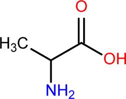
http://www.bmrb.wisc.edu/metabolomics/mol_summary/?molName=D_alanine
pKa of R group: effectively NA
MW: ~90 g/mol
Physiological roles
Converted to beta-alanine and joined with pentoate to give pantothenoic acid. Pantothenoic acid is the core component of Coenzyme-A, one of the most frequently used coenzymes that uses its thioester bond-forming capabilities to drive reactions for numbers of what otherwise would be unfavorable reactions. Here is the breakdown of coenzyme A:
 http://lipidlibrary.aocs.org/lipids/coa/index.htm
To created the strucuture of CoA alanine units need to be converted to beta-alanine, which is a disjointed form of the amino acid, with the amino group on the beta carbon as opposed to the 'classical' alpha carbon.
 http://chembase.com/image_structures-00000001_00025000-13351.png
Bacterial synthesis of beta-alanine uses L-aspartate as shown here:


To created the strucuture of CoA alanine units need to be converted to beta-alanine, which is a disjointed form of the amino acid, with the amino group on the beta carbon as opposed to the 'classical' alpha carbon.

Bacterial synthesis of beta-alanine uses L-aspartate as shown here:

From there, beta-alanine can be incorporated into this very important compound (there are different enzymes that catalyze this reaction in eukaryotes).
There are beta-alanine analogs that are thought to be good potential chemotherapeutics. Details to come. . .
There are beta-alanine analogs that are thought to be good potential chemotherapeutics. Details to come. . .
Alanine is ‘best known’ in the microbial world for its dipeptide bond formation of D-ala-D-ala at the end of the pentapeptide extension from N-acetylmuramic acid (NAM) residues of peptidoglycan polymers. This bond is used for the tetrapeptide linkage of opposite facing NAM residues. L-alanine is synthesized and alanine racemase converts it to its stereoisomer, D-alanine. Two D-alanines are then joined by their respective ligase.

The R groups in this case are -CH3 (methyl) groups, and though chirality is not shown, in this case, the configurations are both D. (Need a refresher on chirality? Here is some help with the CORN rule: http://dwb4.unl.edu/Chem/CHEM869K/CHEM869KLinks/www.ccp14.ac.uk/ccp/web-mirrors/llnlrupp/Xray/tutorial/protein_structure.htm )
The peptidoglycan transpeptidase enzyme (also called Penicillin Binding Protein (PBP) because it will covalently bind beta-lactam rings characteristic of penicillins) will covalently link two bridges (the bridges are present in gram positive bacteria, but between gram-negatives there is no bridge between the tetrapeptide extenstions from NAM units). Here is a good summary of the polymerization of peptidoglycan and cross-linking:

Also, interesting is the fact that tmRNA used in trans-translation is only charged with alanine. Details about how alanine-charged tmRNA will disassemble stalled ribosomes:
http://129.123.92.202/biol5190/PDFs/annurev_tmRNA.pdf
http://129.123.92.202/biol5190/PDFs/annurev_tmRNA.pdf
Synthesis
Most often alanine is synthesized by the reductive amination of pyruvate, but there are other enzymes for which there is support of their function in alanine synthesis. Just in Escherichia coli, there are likely two other pathways that are used to add to the alanine pool. The most supported enzyme is a glutamate-pyruvate aminotransferase, that uses the common alpha amino donor, glutamate to add an amino group to pyruvate (this is the same as what occurs in eukaryotes)

Directly from an another amino acid, another pathway does exist in E. coli,
Bacillus subtilis, and Homo sapiens. This pathway does not aminate a three carbon compound (pyruvate) but will desulfonate a cysteine. To visualize this look at the structure of cysteine.
 http://ecocyc.org/META/NEW-IMAGE?type=PATHWAY&object=PWY0-1021&detail-level=2

Directly from an another amino acid, another pathway does exist in E. coli,
Bacillus subtilis, and Homo sapiens. This pathway does not aminate a three carbon compound (pyruvate) but will desulfonate a cysteine. To visualize this look at the structure of cysteine.

Wednesday, April 18, 2012

Tryptophan
 |
| http://biopsychiatry.com/tryptophan/index.html |
Abbreviation: Trp or W
Molecular Formula: C11H12N2O2
pKa: 2.38 (carboxyl), 9.39 (amino)
Tryptophan is encoded as the codon UGG. Only the L-stereoisome is used in enzyme and structural proteins. D-Trp is occasionally found in some naturally produced peptides. Tryptophan is essential. If it is not provided in growth media, tryptophan must be synthesized by the bacterium.
Synthesis
 |
| http://wickershamsconscience.wordpress.com/2011/11/24/trypytophan-turkey-and-post-prandial-drowsiness/ |
The Trp operon regulates the production of tryptophan. There are several methods for the regulation of tryptophan. The first method is negative transcriptional regulation by TrpR repressor. In this method, tryptophan is the effector (or co-repressor).
 |
| http://bio1151.nicerweb.com/Locked/media/ch18/trp_operon.html |
The second method is translational based-transcriptional attenuation. In this method, transcription terminates in the trp operon before the RNA polymerase can transcribe the first structural gene of the trp operon. Termination occurs within the trpL leader region. This region includes four short sequences designated 1-4. These sequences are partially complementary and hairpin loops can form. Three different hairpins are possible: 1-2, 2-3, and 3-4. The 3-4 hairpin is a transcription terminator. Once this hairpin forms, RNA polymerase will disassociate from the DNA and transcription will not occur.
The leader region contains two adjacent tryptophan (trp) codons. In the absence of tryptophan, the ribosome will stall at one of the two trp codons. This blocks sequence 1and prevents the formation of the 1-2 hairpin. The 2-3 hairpin (anti-termination hairpin) then forms. Formation of the 2-3 hairpin prevents the formation of the 3-4 termination hairpin. This allows the RNA polymerase to transcribe downstream genes. In the presence of tryptophan, the ribosome does not stall at the trp codon. This allows the 3-4 hairpin to form. The ribosome translates the entire leader peptide sequence and stalls at the terminator hairpin. This prevents downstream genes from being transcribed.
This method is possible because prokaryotes begin translating mRNA while the RNA polymerase is still transcribing the DNA sequence. For more information, please read trp RNA-Binding Attenuation Protein (TRAP)-trp Leader RNA Interactions Mediate Translation as well as Transcriptional Regulation of the Bacillus subtilis trp Operon by Enrique Merino, Paul Babitzke, and Charles Yanofsky.
 |
| http://www.nature.com/scitable/content/two-different-secondary-structures-may-be-formed-19425 |
Tyrosine

Abbreviated: Tyr or Y
Molecular formula: C9H11NO3
pKa: 2.20(carboxyl), 9.11(amino), 10.07(R Group)
Physiological Roles
The R group of tyrosine is polar. Its codons are UAC and UAU. Tyrosine is classified as a non essential amino acid, it can be synthesised. But if you want more in your diet though tyrosine can be found in high protein foods such as chicken, fish, peanuts, milk, cheese and yogurt.
Tyrosine is plays important roles in the nervous system. One of its fates is that it is converted into catecholamines. Catecholamines are molecules that have a catechol nucleus consisting of benzene with two hydroxyl side groups, and a side-chain amine. In the nervous system the catecholamines are the always important dopamine, norepinephrine, and epinephrine, all neurotransmitters. (King 2012)

This occurs by tyrosine first being converted into DOPA (3,4-dihydrophenylalanine) by a tyrosine hydroxylase. From there a DOPA carboylase will convert DOPA into dopamine which in turn can be synthesised into norepinephrine and epinephrine.
In bacteria protein-tyrosine kinases have been found to phosphorylate protein substrates, including RNA polymerase sigma factors, UDP-glucose dehydrogenases and single-stranded DNA-binding proteins, all important in cellular life (Mijakovic et al). Because of this particular activity new antimicrobial drugs are being made to target the tyrosine kinases and the phosphorylation of certain endogenous proteins catalyzed by specific tyrosine kinases (Cozzone).
Synthesis

Tyrosine synthesis begins with a chorismate. The chorismate is converted to prephenate by chorismate pyruvatemutase.
From there the prephenate is acted on by prephenate dehydrogenase (NADP+)and NAD+ to make a carbon dioxide, NADPH and 4-hydroxyphenylpyruvate.
The final step occurs when L-glutamate and 4-hydroxyphenylpyruvate are acted on by a tyrosine transaminase to make 2-oxoglutuate and L-tyrosine (Enzyme Database)
Subscribe to:
Posts (Atom)